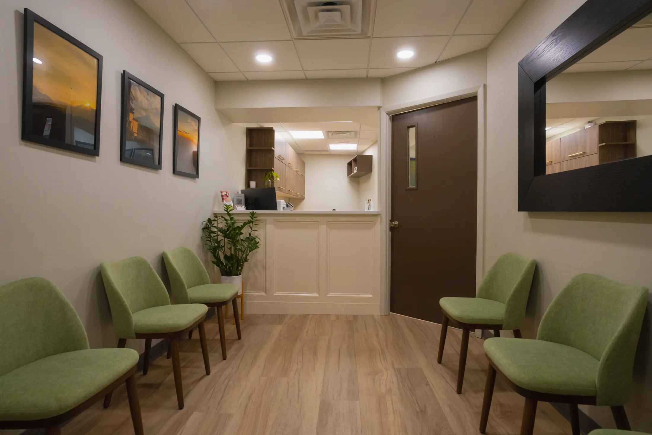
Inaba Dental Office
Family centric dentistry in English and Japanese for Westchester and Long Island.
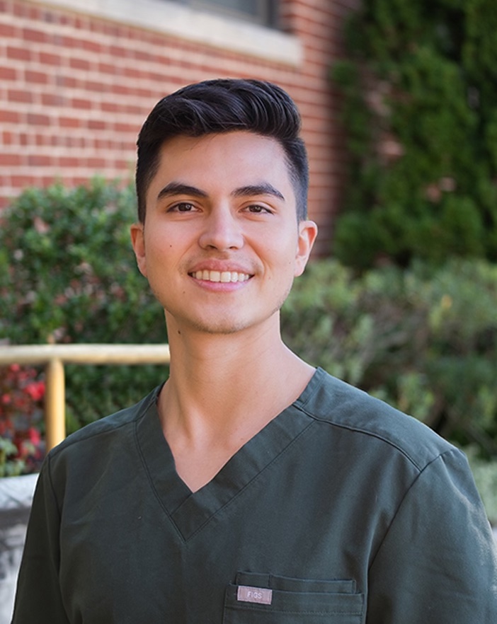
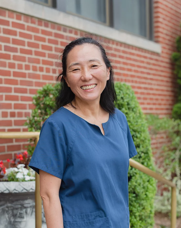
Dr. Takekazu Inaba opened Inaba Dental in 1990. Today, his daughters; Dr. Rie and Dr. Rina Inaba, along with their husbands, Dr. Matthew Rhee and Dr. Joshua Penners continue to expand and improve the practice and services offered in order to provide the highest quality dental care for our patients.
A family dental practice
The same family of doctors for two generations, and the same front desk that has welcomed patients since the practice began

Services
With multiple doctors in one practice, we are able to offer a variety of services to meet the needs of our patients
Prevention & diagnostics
Exams, cleanings, digital X-rays, 3D imaging, and oral-cancer screening — the visits that find problems while they are still small.
Restorative dentistry
Same-day CEREC crowns, inlays, and onlays milled in our office, plus bridges, fillings, and removable options.
Endodontic treatment
Emergency relief for pain and swelling, primary root canals, retreatment of failing canals, and apicoectomy when a root tip needs surgery.
Periodontal (gum) care
Gum disease treated at every stage, from deep cleanings to surgery, and soft-tissue grafting to reverse recession.
Dental implants
A titanium post replaces the missing root. Bone grafting, sinus lifts, and site preparation are planned and performed here.
TMJ, grinding & clenching
Custom night guards, therapeutic Botox, treatment of worn teeth and the bite, and management of jaw pain.
Scarsdale, Westchester
2 Overhill Road, Suite #110, Scarsdale, NY 10583
By appointment
Westbury, Long Island
265 Post Avenue, Suite #110, Westbury, NY 11590
By appointment




 U.S. Department of
Veterans Affairs
Professional organizations our doctors belong to.





“Despite my tight and unbending schedule due to leaving the country for work, Dr. Inaba expedited and prioritized my treatment. She was gentle with my teeth and provided clear instructions on how to proceed. A family practice that conducts themselves with genuine care and integrity.”
“They cared for me during my time posted here. At first I was scared of getting an implant, but the warm, family-like staff helped me and before I knew it my fear had melted away. In an unfamiliar life abroad, I was truly glad I could receive treatment using the latest equipment and techniques.”
“The atmosphere in the office is so warm, and the doctors and front-desk staff are all so kind that my son — who hates the dentist — was able to come back again and again without any fuss. Thank you so much!”
Services
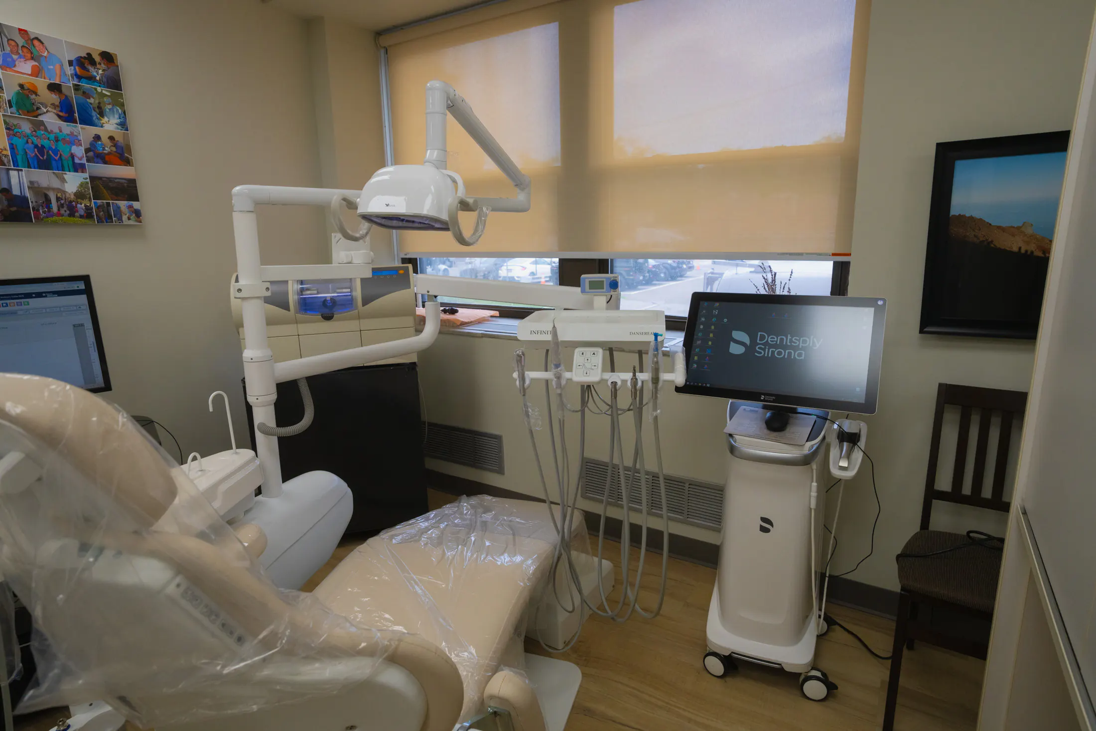
Our doctors have invested in the education, equipment and technology to be able to offer a wide variety of cutting edge dental services.
On this page you'll find more details about the services we provide. Your treatment plan will be made and reviewed with you during your examination to ensure you recieve the correct diagnosis and treatment recommendations.
Preventive care
Exams & check-ups
Full examination, extraoral photos, digital X-rays, non-invasive cavity detection aid, and oral-cancer screening.
Dental cleanings
Professional cleanings every 3–6 months depending on your gum health.
Gum-disease treatment
Deep cleanings (scaling & root planing) with local anesthetic for comfort.
Night guards
Custom guards to protect teeth and jaw joints from grinding and clenching.
Botox for TMJ & clenching
Therapeutic Botox to relax overactive jaw muscles, easing TMJ symptoms, bruxism, clenching, and related jaw pain.
Pediatric dentistry
First visit by age one; sealants, fluoride, fillings, and gentle habit guidance.
Interceptive orthodontics
Early appliances that guide jaw growth and can shorten future braces or aligner time.
Sealants
A thin protective coating over deep grooves to keep cavities from starting.
Silver diamine fluoride
A no-drill liquid that stops early decay, ideal for young children.
Restorative dentistry
Tooth-colored fillings
Natural-looking composite fillings; ICON resin infiltration for white spots without drilling.
Same-day crowns (CEREC)
Digitally designed ceramic crowns, inlays, and onlays milled in our office. No temporary crown needed.
Bridges
A replacement of one or more missing teeth anchored to neighboring teeth.
Extractions
Removing a tooth that cannot be saved, often with bone grafting when planning an implant, and immediate implant replacement when possible.
Wisdom tooth removal
Removing crowded or impacted wisdom teeth, planned on a Cone Beam 3D scan.
Root canal treatment
A way to clear an infection while preserving your natural tooth.
Dental implants
A dental implant replaces the root of a missing tooth with a small titanium post that fuses with the bone, giving a crown a stable, natural-feeling foundation.
Dentures
Partial and full dentures, including implant-stabilized options.
Diagnostic technology
Cone Beam 3D imaging and Spectra fluorescence cavity detection.
Periodontal (gum) care
Treatment of periodontal disease
From deep cleanings to surgical care — cleaning roots directly, reshaping damaged bone, and regrowing lost support with grafts and membranes to save the tooth.
Soft-tissue (gum) grafting
Periodontal plastic surgery to reverse gum recession and ensure health and longevity to your smile.
Crown lengthening
Reshaping the gumline to expose enough healthy tooth for a crown to hold onto.
Socket preservation
A graft placed the day a tooth is removed keeps the socket from collapsing, so the site stays ready for an implant.
Ridge augmentation
Rebuilding width and height where the jaw has shrunk after years without a tooth, giving an implant solid ground.
Sinus lift
For upper back teeth: gently raising the sinus floor and adding bone beneath it to create the height an implant needs.
Gum grafting around implants
Thickening the collar of gum that seals around an implant, for a healthy, natural-looking result.
Cosmetic dentistry
Teeth whitening
In-office whitening or custom take-home trays.
Veneers
Thin porcelain facings for stained, uneven, or misshapen front teeth.
Clear aligners (Invisalign)
Discreet straightening that also improves your bite.
Gap & black-triangle closure
The Bioclear injection-molding method closes spaces with little to no drilling.
Botox
Softens expression lines, and can relax a jaw that clenches or a gummy smile.
Replacement option for one or more missing teeth.
A dental implant is a small titanium post that is placed in the jaw. After a period of integration, an implant crown or bridge can be made.
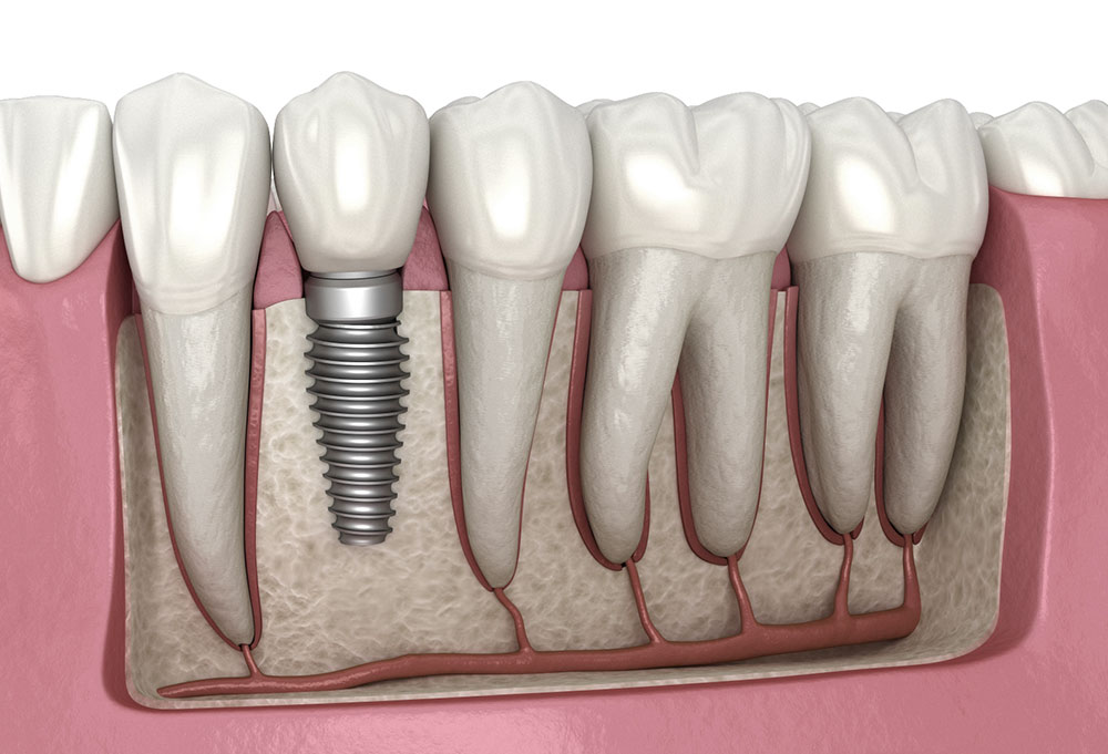
Illustration: Alexmit art · CC BY-SA 4.0
What is a dental implant?
Unlike a bridge, an implant doesn't require preparing the neighboring teeth. The treatment can be isolated to only the affected tooth or teeth. After a period of healing, termed osseointegration, the implant is ready for use. An implant crown can then be attached to the implant, usually via a small screw. The implant and implant crown are designed to handle the forces of eating, chewing, and daily use.
Oftentimes, dental implants can be placed at the same time that a tooth is extracted. Other times, bone grafting may be required prior to dental implant placement. For anterior teeth that show in the smile, sometimes a temporary crown can be made at the same time as the implant placement, providing a temporization solution that does not involve a removable appliance.
The procedure
Every case is different, but most follow these stages.
Consultation and imaging
We review your health history and take any imaging required to accurately diagnose and treatment plan your case. Imaging often includes 3D cone beam computed tomography.
Surgical phase
The surgical phase of the treatment plan is tailored to your specific situation. This phase will include the placement of the dental implant, but may also require bone grafting, soft tissue grafting, ridge augmentation, tooth extraction, or sinus lift procedures.
Healing
After implant placement, a healing period is required for bone to fuse around the dental implant. This process is termed: osseointegration. It is typically 3-4 months depending on the clinical situation.
Custom crown fabrication
After an impression appointment, a customized restoration is designed and fabricated for the dental implant. The design is carefully considered for longevity, cleansibility, aesthetics, and comfort.
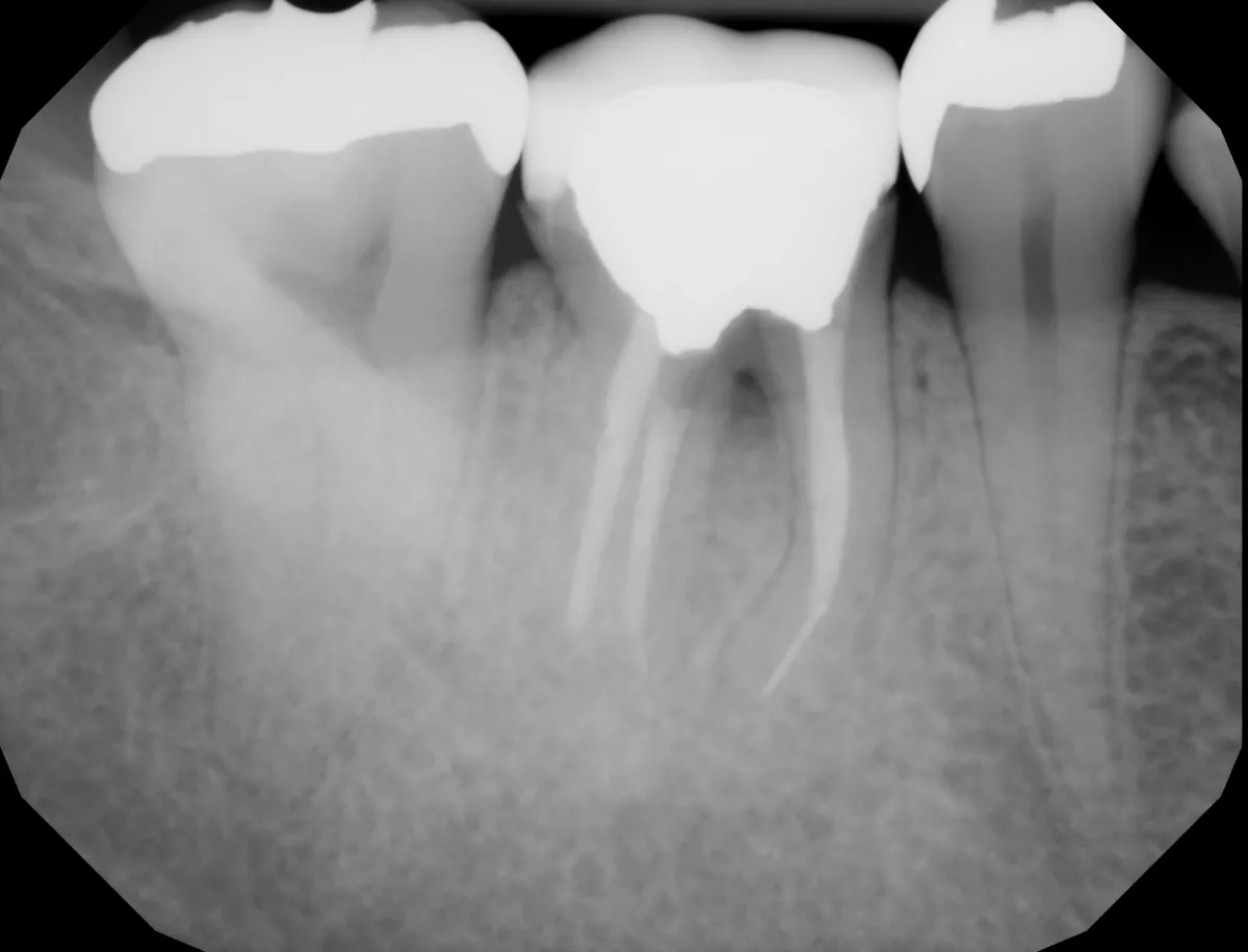
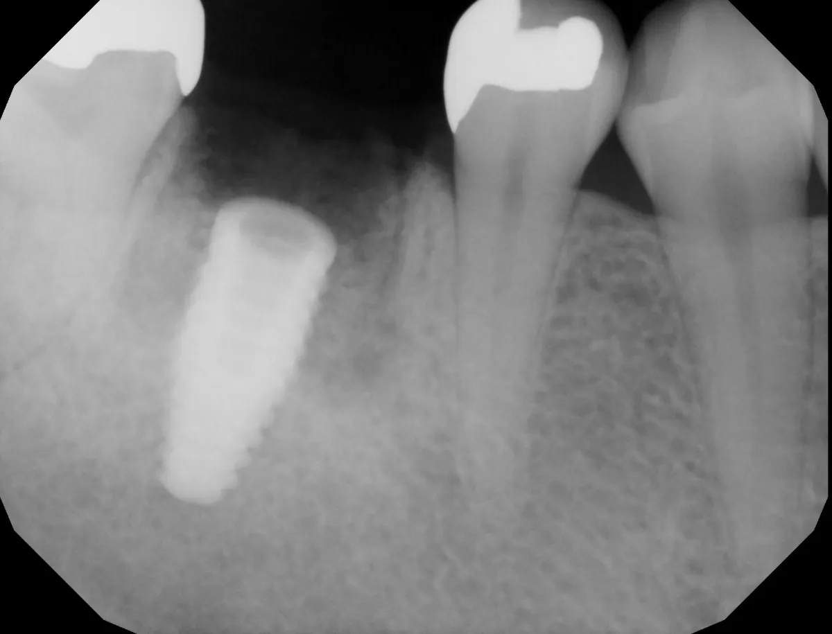
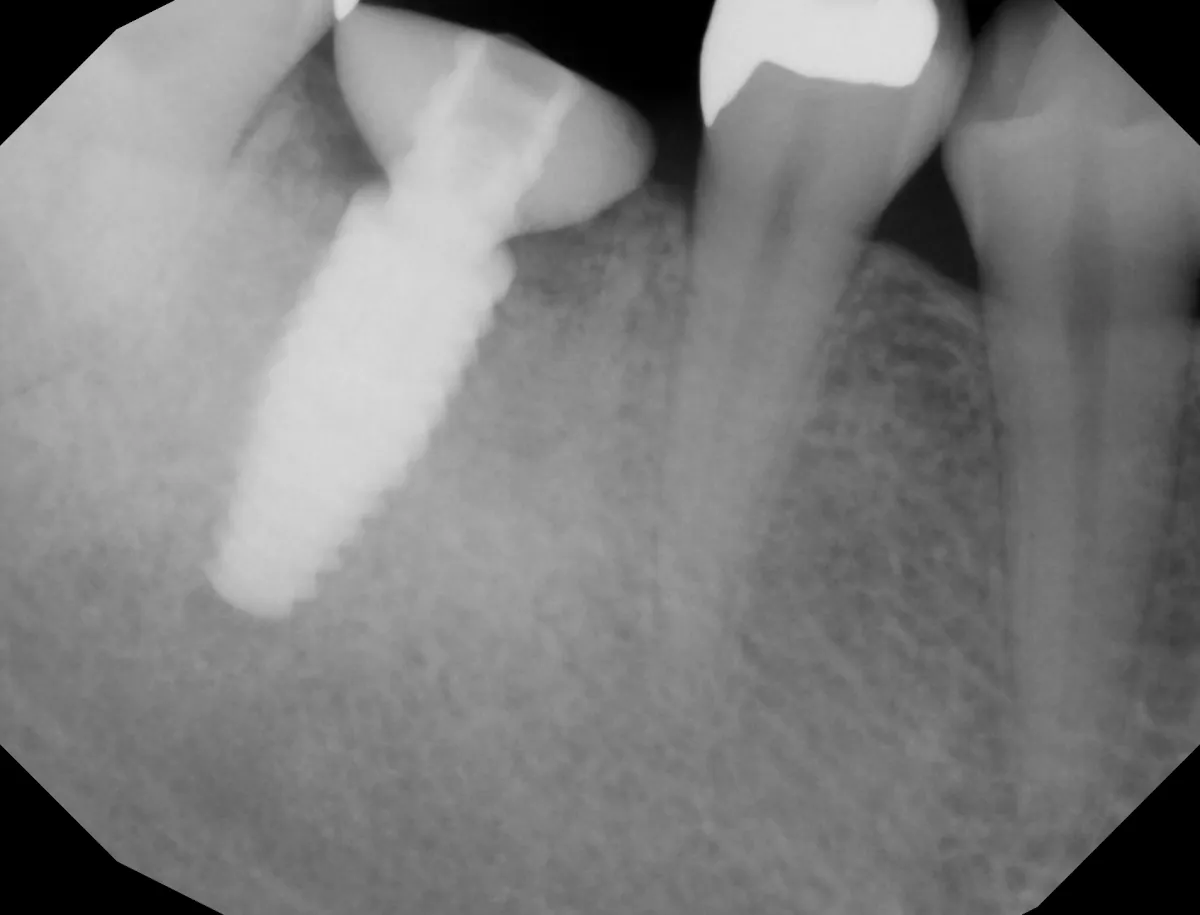
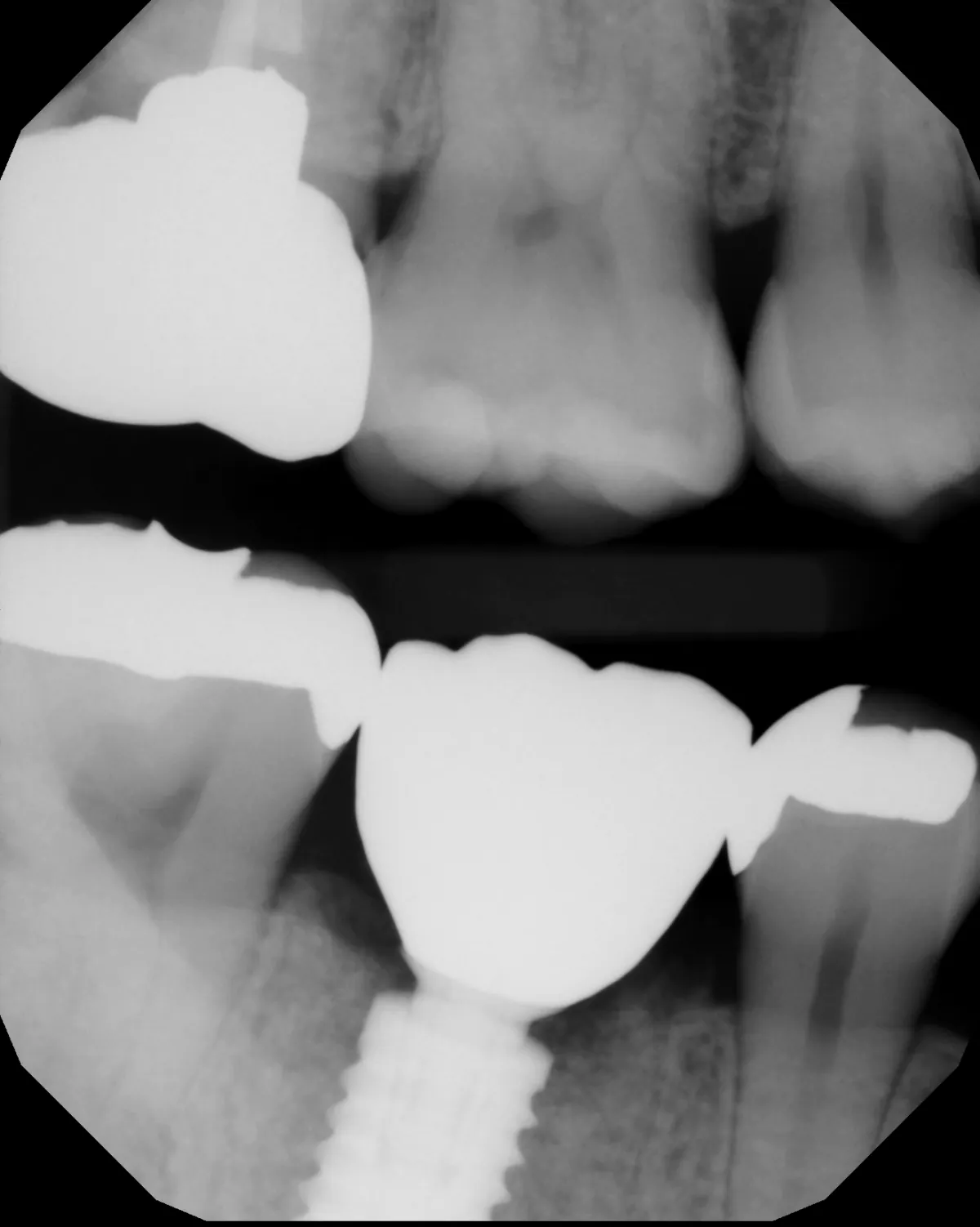
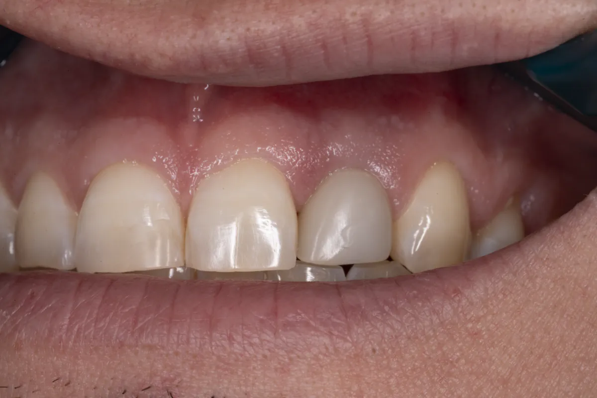
Immediate implant — posterior, with custom healing abutment
Sometimes, a dental implant can be placed at the same time as tooth extraction. When paired with a bone graft and a customize healing abutment, this treatment option helps to significantly shorten the treatment duration as well as ensure that the gum architecture is preserved for healthy, long-term restoration.
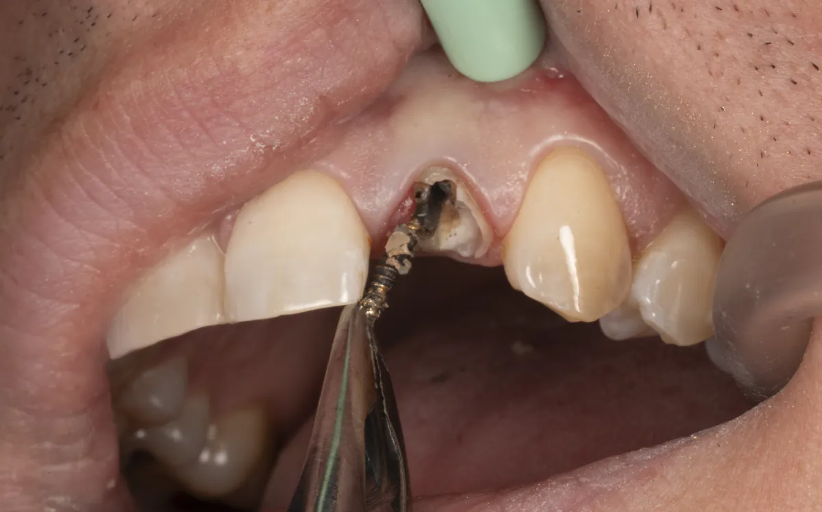
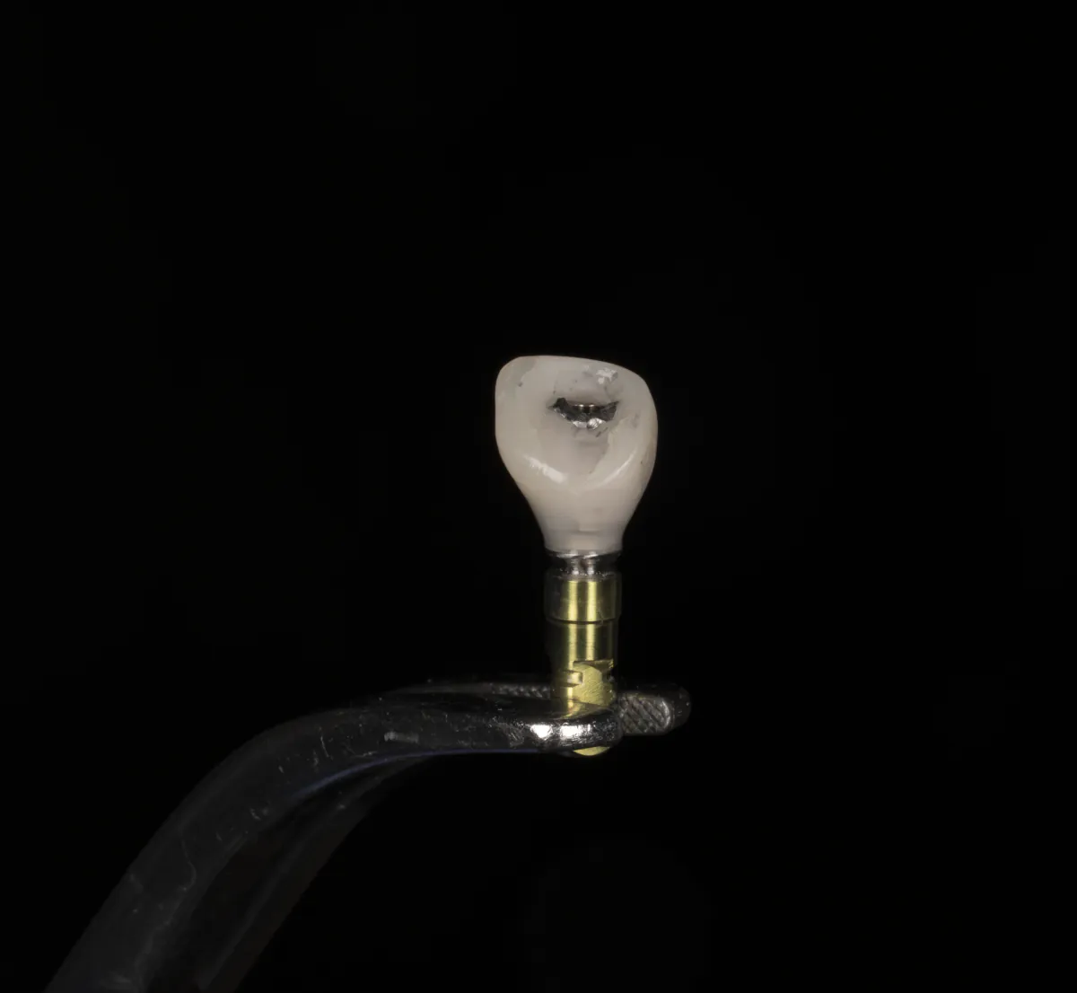
Immediate implant — anterior, with provisional crown
For front teeth that show in the smile, an immediate implant can often be paired with a provisional crown made the same day. The temporary crown holds the shape of the gums to prevent any collapse, and a removable temporary denture is avoided.
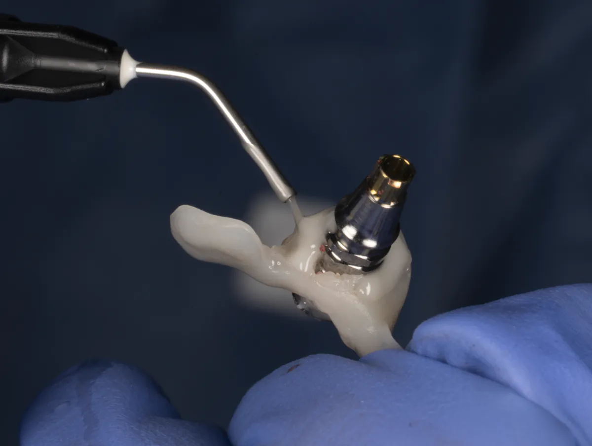
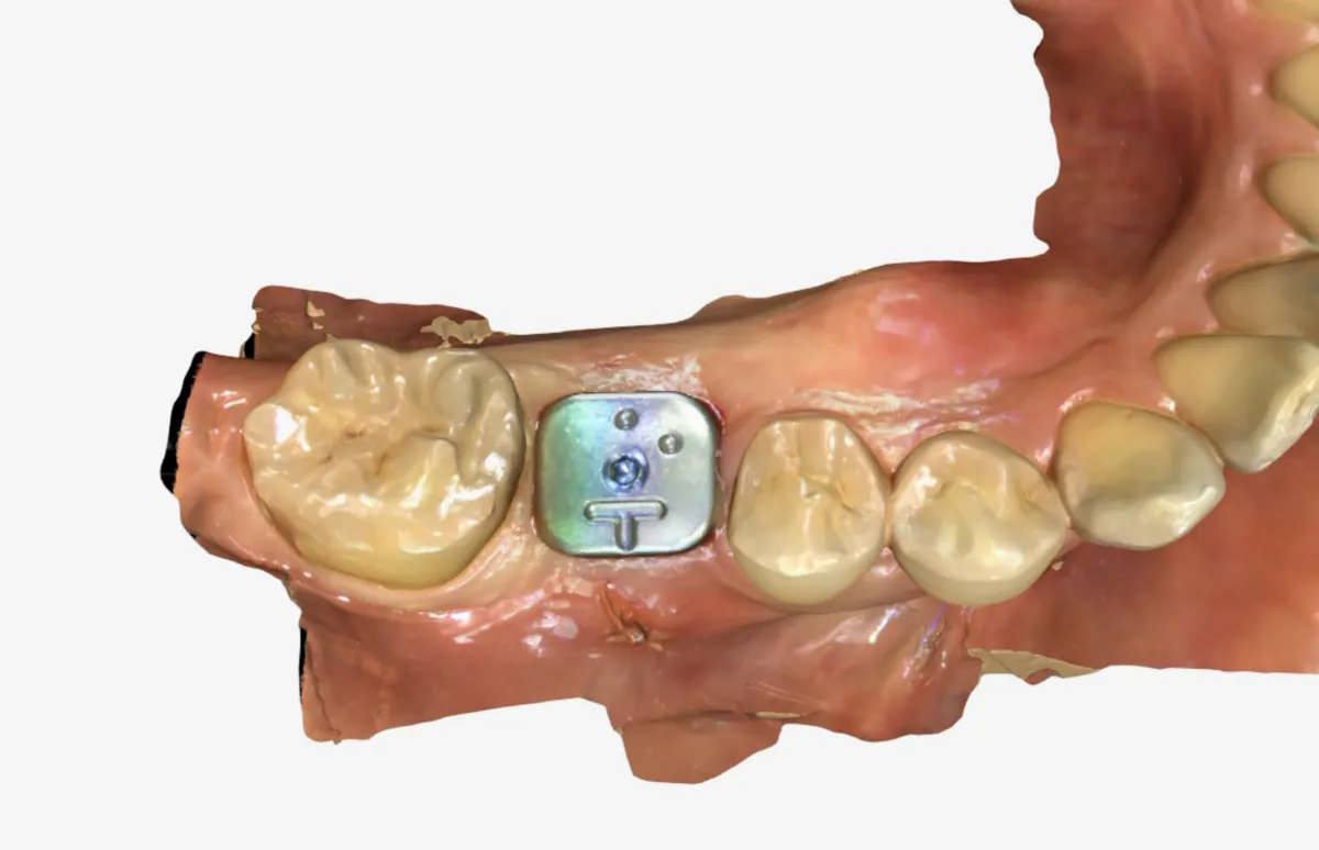
Delayed implant placement — with scan healing abutment
When the site needs time to heal first, the implant is placed once the bone has matured, with a scan healing abutment that shapes the gum and doubles as a digital scan reference. After about 3 months of healing, a custom abutment is fabricated to support the final crown.
Implant placement after grafting — GBR, sinus lift, and soft-tissue grafts (CTG / FGG)
When bone or gum has been lost, the site is rebuilt before or alongside implant placement: guided bone regeneration (GBR) to restore bone volume, a sinus lift for the upper back jaw, and soft-tissue grafting — connective tissue (CTG) or free gingival (FGG) grafts — to thicken and strengthen the gum around the implant.
PLACEHOLDER: grafting case sequences (GBR, sinus lift, CTG, FGG), to be added
Procedures that often go with an implant
Questions patients ask
Does getting an implant hurt?
The procedure itself is done under local anesthesia. A pain protocol will be customized to you along with your post operative instructions. Most simple implant surgeries have very little post operative pain and swelling.
How long does the whole process take?
Your treatment plan will be made at the initial consultation and an estimate of the total time will be presented. An implant treatment plan can range anywhere from 3 months if the impression is taken at the time of surgery or up to 12 months if procedures such as ridge augmentation or sinus augmentation are required.
Will the implant look natural?
We take photos to document the shade (color) of your existing teeth, and communicate with our dental laboratory. In some cases, we also stain and glaze our ceramic restorations in house for a custom solution.
Same day ceramic restorations
CEREC CAD/CAM is a way for a dentist to make a crown (or inlay/onlay) right here in the office, in a single visit, using a computer and a milling machine.
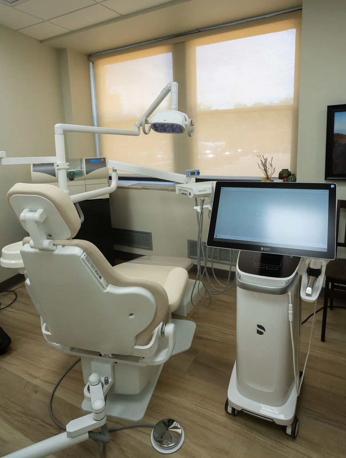
What is CEREC?
Instead of packing a tray of impression putty over your teeth, a small camera builds an accurate 3D model on the screen. From there we design the crown, inlay, or onlay right on that model, adjusting the bite, the way it meets the teeth on either side, and the overall shape before anything is actually made.
The milling unit then carves the restoration out of a solid ceramic block. That usually takes about 6 to 15 minutes. Compare that to a traditional lab crown, which takes two visits two to three weeks apart and a temporary crown to get you through the wait. With CEREC it all happens in one appointment – no second visit, no temporary crown, no waiting for weeks. You walk out the same day with the final restoration.
The procedure
One appointment, usually about 1.5 to 2 hours.
Preparing the tooth
After numbing the area, the cavity or old filling is removed and the tooth is shaped to give the new restoration a precise fit.
Digital scan
Instead of the putty mold, a small wand-like camera captures the newly prepared tooth and creates a precise 3D picture of it on a screen. No impression tray, no putty.
Design and milling
The software designs a custom restoration that fits your tooth exactly, like designing a puzzle piece to fill a specific gap. That design gets sent to a milling machine that takes a solid block of ceramic and carves the restoration out of it, which is then polished (and sometimes stained and glazed if in a very aesthetic area) and cemented.
A same-day CEREC crown, start to finish
A typical case: a tooth with a large, failing filling needs full coverage. The photos below follow one tooth through a single visit, before, during, and after.
Treatments that may go with a crown
Questions patients ask
Do I still need a temporary crown?
Most of the time no. In rare instances the tooth may be temporized to monitor for symptoms.
Is a same-day crown as strong as a lab-made one?
For everyday crowns, inlays, and onlays, CEREC restorations are strong, precise, and long-lasting. For the most cosmetically demanding cases, or implant crowns or multi-tooth bridges, a traditional lab is sometimes still preferred for custom shading; we will discuss with you which approach suits your situation.
Will it look natural?
Yes. We choose the ceramic block against your natural teeth and then stain and glaze if necessary.
What does the scan feel like compared to the old putty impressions?
Much easier. Instead of biting into a tray of impression material, a small camera captures photos of the tooth to create a 3-dimensional scan.
How long will it last?
With proper care, a well-fitted ceramic restoration can last many years, comparable to a traditional crown. Keeping up with brushing, flossing, and regular checkups makes a real difference.
Treatment to save a damaged tooth.
A root canal removes infected or inflamed tissue from inside the tooth. Once cleaned and sealed the tooth can be restored and kept for years to come.
What is a root canal?
Unlike an extraction, a root canal keeps your natural tooth in place. The treatment targets only the soft tissue, or pulp, inside the tooth. When a cavity or trauma causes irreversible damage to the pulp, it can become infected and painful. During the root canal procedure, this diseased tissue is removed, cleaned, and disinfected.
After cleaning, the canals are filled and sealed to prevent reinfection. Because a treated tooth can become brittle over time, a crown is usually placed afterward to protect it and restore full chewing strength. The tooth and its restoration are designed to handle the everyday forces of eating and daily use.
The procedure
Every case is different, but most follow these stages.
Consultation and imaging
We review your health history, examine the tooth, and take any imaging required to accurately diagnose the problem and plan your treatment.
Cleaning and shaping the canals
After numbing the area, a small opening is made in the tooth. The infected or inflamed pulp is removed, and the canals are carefully cleaned, shaped, and disinfected. This step may take multiple visits depending on the tooth.
Filling and restoration
The cleaned canals are filled and sealed to prevent reinfection. Because a treated tooth can become brittle, a crown is usually placed afterward to protect it and restore full chewing strength. Healing is typically quick, with any tenderness easing over a few days.
Endodontic retreatment — molar, persistent infection
Sometimes a tooth that had a root canal fails to heal or becomes reinfected years later. In retreatment, the previous filling material is removed, the canals are re-cleaned and disinfected, and the tooth is resealed — often saving it without surgery and preserving the natural tooth long-term.
Apicoectomy — root-end surgery on a re-infected tooth
When infection persists at the tip of the root and conventional treatment isn't enough, an apicoectomy removes the very end of the root and the surrounding infected tissue through a small opening in the gum. A tiny filling seals the root end, allowing the bone to heal while the tooth stays in place.
Internal bleaching — discolored front tooth after root canal
A tooth can darken after trauma or a root canal, as the tissue inside it changes color. Internal bleaching places a whitening agent inside the treated tooth to lighten it from within, restoring a natural shade without a crown or veneer, and preserving healthy tooth structure.
Procedures that often go with a root canal
Questions patients ask
Does a root canal hurt?
The procedure itself is done under local anesthesia, so most patients feel some pressure. A pain protocol will be customized to you along with your post procedure instructions. Modern root canals are typically comparable to having a routine filling placed.
How long does the whole process take?
Your treatment plan will be made at the initial consultation and an estimate of the total time will be presented. Many root canals are completed in a single visit, while more complex cases or teeth with multiple canals may require two appointments. A crown is usually placed in a later visit to protect the tooth.
Will the tooth look natural afterward?
Yes. Once the tooth is sealed, it is restored with a filling or a crown that is color-matched to your existing teeth for a natural-looking result.
Is a root canal better than pulling the tooth?
In most cases, keeping your natural tooth is the best option. It preserves normal biting and chewing, protects the surrounding teeth from shifting, and avoids the need for an implant or bridge. We help you weigh the options for your specific situation.
Soft Tissue Grafting
When the gum recedes, the root of the tooth is left exposed: it is softer than enamel, more sensitive to cold, and easier for decay to reach. A soft-tissue graft covers the root and thickens the gum so the recession does not keep advancing. Implants rely on the same firm collar of gum, and the same techniques build it.
Why gums recede
Recession usually has more than one cause, and knowing yours shapes the treatment. The most common:
Periodontal disease
Infection breaks down the bone and gum that hold the tooth. Treating the disease comes first; grafting rebuilds what was lost.
Brushing too hard
A stiff brush or a heavy scrubbing stroke wears the gum away over years, often on the canines and premolars.
Naturally thin tissue
Some people inherit a thin gum type that recedes more easily. Thickening it is often the main goal of the graft.
Tooth position
A tooth that sits outside the arch, or moved there during orthodontics, can end up with very little gum and bone on its outer surface.
Grinding and clenching
Heavy bite forces stress the thin bone over the root. A night guard often accompanies grafting so the result holds.
Muscle and frenum pull
A tight band of tissue between lip and gum can tug the gumline down. Releasing it is sometimes part of the same procedure.
Where the graft tissue comes from
There are two sources, and we choose between them with you at the planning visit. Both are performed here in the office.
Harvested from the palate
A thin layer of connective tissue is taken from under the surface of the roof of your mouth and placed at the recession site. Because the tissue is your own, it integrates predictably and gives the most durable root coverage; it remains the gold standard, especially for one or two teeth.
The trade-off is a second surgical site: the palate feels like a pizza burn for about a week while it heals, and the amount of tissue we can take in one visit is limited.
Prepared donor tissue (allograft)
Sterile, medically processed donor tissue serves as a scaffold that your own cells grow into. Nothing is taken from your palate, so there is only one surgical site, recovery is more comfortable, and we can treat many teeth in a single visit.
Root coverage is very good, though on average slightly less thick and slightly less stable over decades than your own tissue. We often recommend it when several teeth need grafting at once, or when the palate is thin.
Grafting around implants
An implant cannot get a cavity, but the gum around it still decides how long it lasts. We usually graft at the time of implant placement or uncovering, so everything heals at once; an implant already in place can be grafted too.
A seal that protects the bone
A band of keratinized tissue, the firm pink gum that resists rubbing, seals the implant off from food and bacteria. Thin, loose tissue lets inflammation reach the bone that holds the implant.
Comfortable daily cleaning
Grafted tissue does not shift when you chew or brush, so the site stays comfortable and easy to keep clean. That daily cleaning is what keeps an implant healthy for decades.
A natural-looking gumline
For front teeth, grafting thickens the collar of gum so the implant crown emerges the way a natural tooth does, with no gray shadow at the edge.
The procedure
A single visit of about 60 to 90 minutes, under local anesthesia.
Exam and planning
We measure the recession, check the bite and brushing habits that caused it, and decide together on palate tissue or donor tissue. If gum disease is active, we treat that first.
Preparing the site
After numbing, the root surface is cleaned and the gum at the recession is gently loosened, often through a small tunnel technique that avoids larger incisions.
Placing the graft
The graft, from your palate or the prepared donor tissue, is tucked under the loosened gum, positioned over the root, and secured with fine dissolvable stitches.
Healing and follow-up
Soft foods and no brushing at the site for the first two weeks; we check healing at one week and again at four to six weeks. The new gumline matures over about three months.
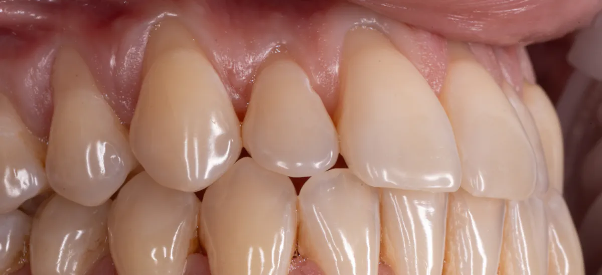
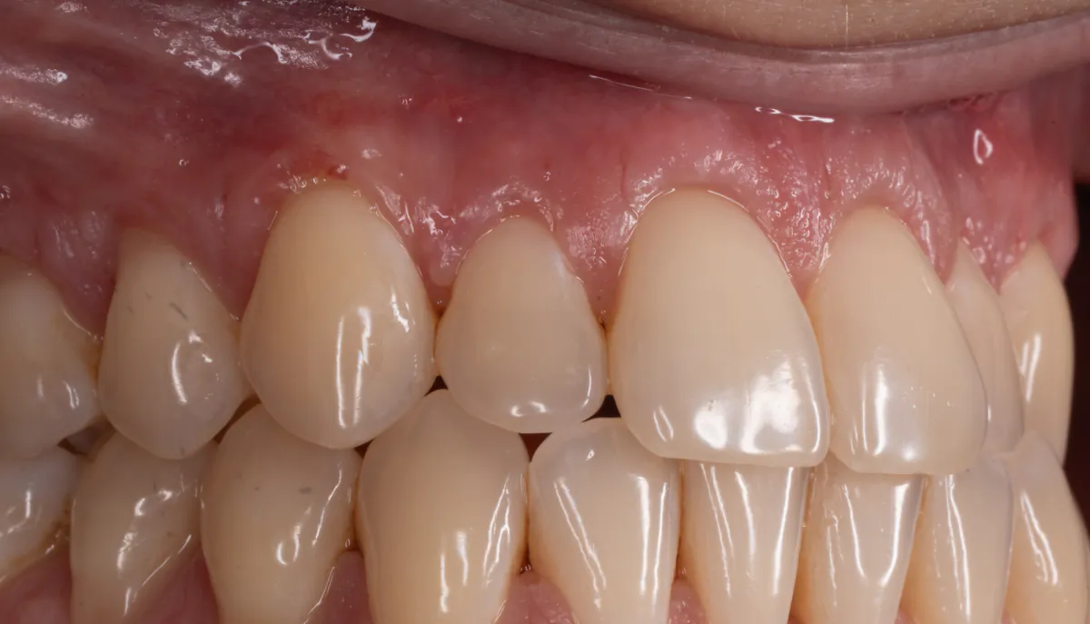
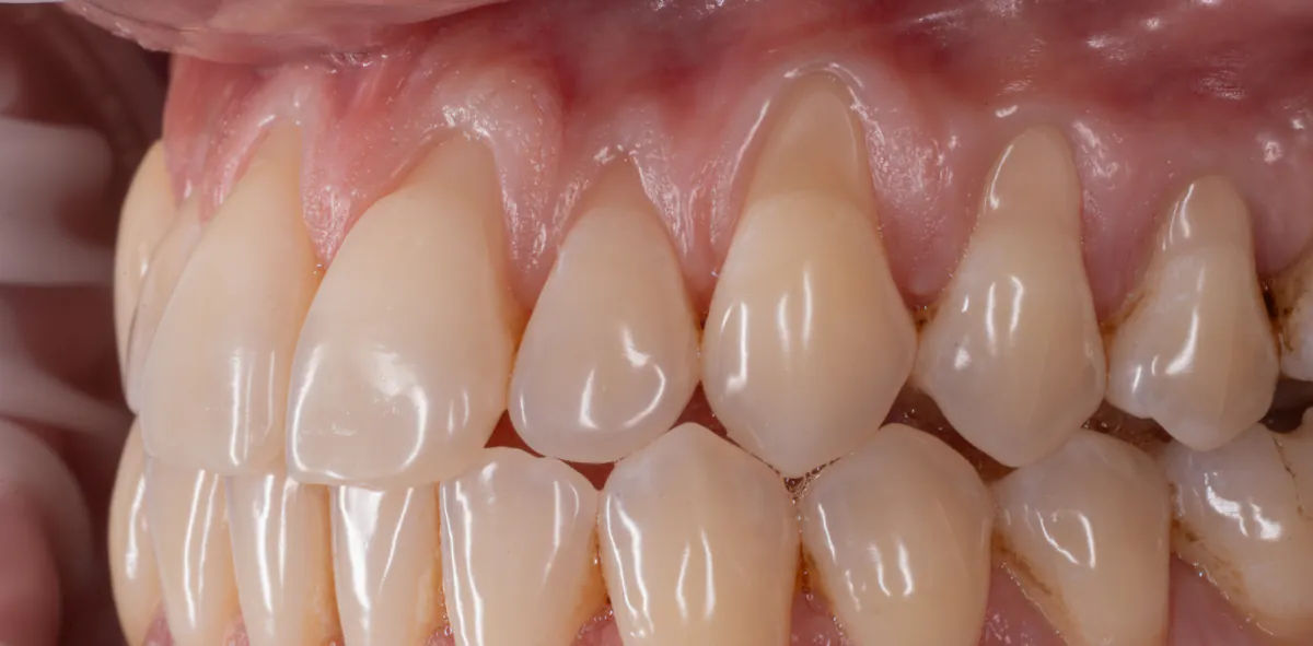
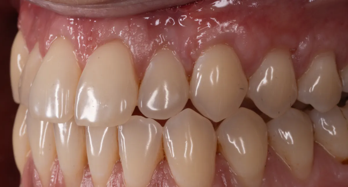
Soft Tissue Grafting using Tunneling Approach
Related care
Questions patients ask
Does gum grafting hurt?
The procedure itself is done under local anesthesia and you should feel pressure, not pain. Afterward, most patients manage with ibuprofen or acetaminophen for a few days. When tissue is taken from the palate, that site is usually the sorer of the two; a simple retainer-like guard can cover it while it heals.
How long is recovery?
Most people are back to work or school the next day. Plan on soft foods for about two weeks and no brushing at the grafted site until we clear it; the gumline continues to mature for about three months.
Will the graft last, or can the gum recede again?
A successful graft is usually stable for decades, provided the cause is addressed: a softer brush and lighter stroke, a night guard if you clench, and regular periodontal maintenance. That is why the planning visit spends as much time on causes as on the surgery itself.
Is donor tissue safe?
Yes. Allograft tissue comes from certified tissue banks regulated by the FDA, and is screened, sterilized, and processed so that no donor cells remain: it acts as a scaffold your own tissue grows into and replaces. It has been used in periodontal surgery for decades.
Treatment Philosophy
Most dental issues, such as tooth decay, gum disease, cracked teeth, and gum recession, are simpler to treat when caught early. Our exams are individualized to you. We take careful records and use modern diagnostic technology to identify and diagnose a variety of dental diseases and conditions. Our treatment philosophy is centered around prevention and risk reduction.
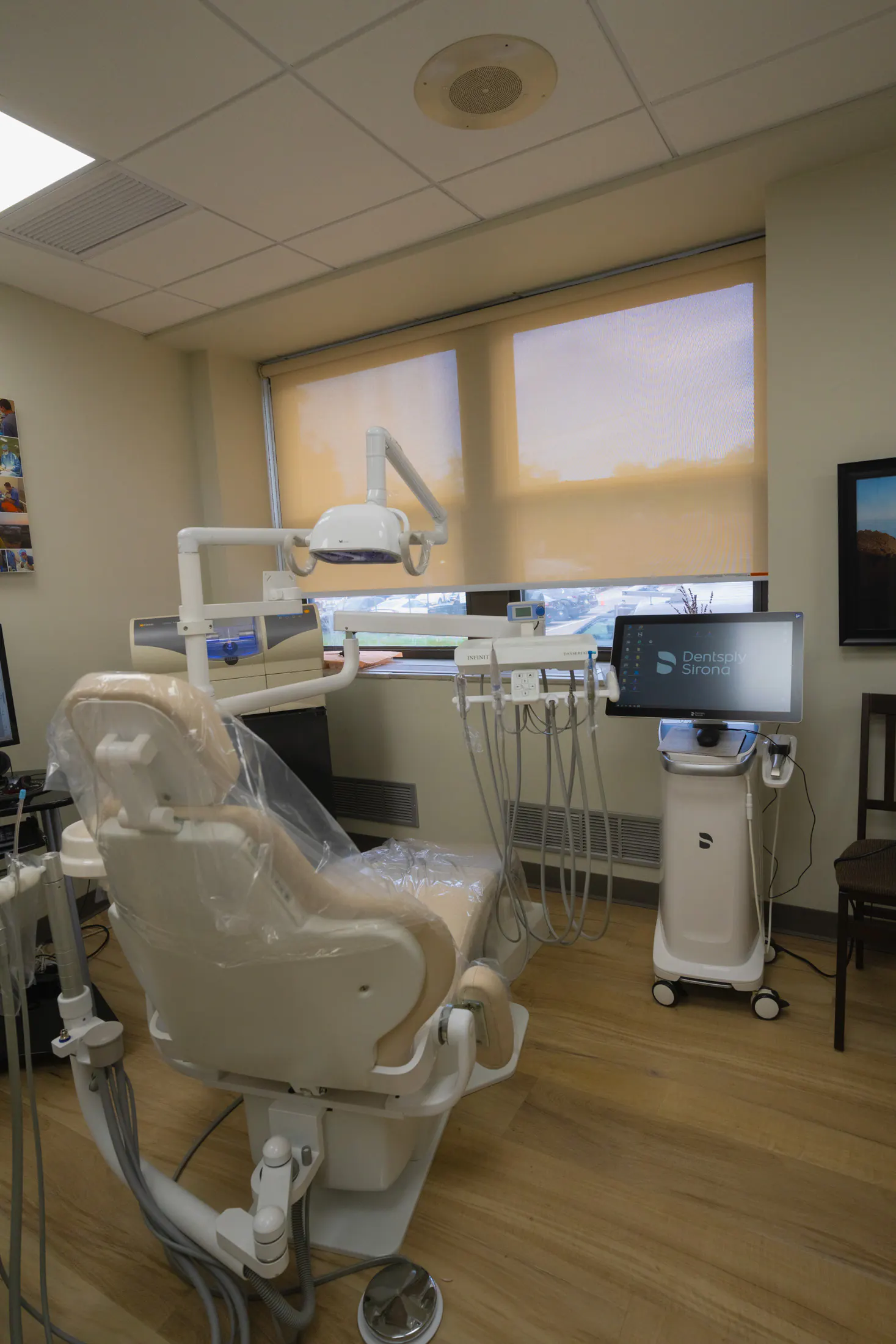
Document, compare, treat conservatively
Dentistry works best when nothing is a surprise. At every exam we build a record — photos, measurements, and imaging when it is needed — and set it beside the records from your previous visits. A groove that is starting to stain, a crack that is growing, a gum line that is receding: change over time tells us far more than any single visit can.
When something does need attention, we start with the most conservative option that works: fluoride, sealants, or watchful monitoring before a drill, whenever the evidence supports it. And because we see whole families — first checkups through grandparents — prevention is tailored to every age.
Diagnostic technology
Each tool answers a different question. Together they give us a complete picture of your mouth — and a record we can compare, visit to visit.
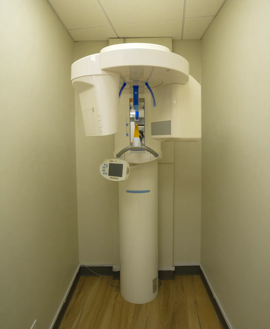
Cone beam CT: 3D imaging, in the office
A conventional X-ray flattens everything into one two-dimensional picture. Our cone beam CT (CBCT) scanner rotates around your head in a few seconds and builds a precise 3D model of the teeth, roots, jawbone, nerves, and sinuses instead.
We use it when the third dimension changes the plan: placing implants precisely, tracing root canals, locating impacted wisdom teeth relative to nerves, and diagnosing pain that flat images cannot explain. Scans happen here — no referral to an imaging center — and we limit the field of view to the area in question to keep the dose low.
New photos at every exam
A fresh set of clinical photos at each visit — not just your first — so changes show up as a series, and we can show you rather than just tell you.
Digital X-rays
High-resolution digital sensors at a fraction of the dose of film, on screen instantly. Taken on a schedule based on your individual risk — not a fixed routine.
Spectra caries detection
A fluorescence camera that makes early decay in deep grooves visible before it shows clearly on an X-ray — no radiation, and a score we can track over time.
Intraoral 3D scanner
A small camera builds a precise digital model of your teeth in minutes — no impression putty. It powers same-day crowns, night guards, and aligners, and tracks wear and movement over time.
Gum measurements
Gentle measurements around each tooth map the health of your gums and bone, so gum disease is caught while it is still reversible.
Oral cancer screening
A check of the tongue, cheeks, lips, and palate for lesions, lumps, or discoloration is part of every exam.
What to expect at your exam
The routine is the same at every visit — so nothing is missed, and everything is compared.
A conversation, and new photos
We start by listening: health changes, concerns, anything that feels different. Then we take a fresh set of photos — at every exam — which become the baseline we compare against next time.
Imaging, only as needed
Digital X-rays are taken at intervals matched to your individual risk, at a fraction of the dose of film. When a case calls for three dimensions, the cone beam CT is just down the hall.
The examination itself
Tooth by tooth for decay, cracks, and wear; the Spectra camera on any suspicious groove; gentle measurements around each tooth to map gum health; and an oral cancer screening.
Review together, then a plan
You see what we see: today's photos and X-rays beside earlier visits, on the screen in front of you. We decide on a plan together — often simply watching — and set the cleaning interval that fits you. Exams usually pair with a cleaning the same day.
Preventive treatments
Small, simple measures now that prevent bigger treatment later.
Dental cleanings
Professional cleanings every 3–6 months depending on your gum health.
Fluoride treatment
Professional fluoride strengthens enamel and can remineralize the very earliest decay — for children and for cavity-prone adults.
Sealants
A thin protective coating over deep grooves to keep cavities from starting.
Silver diamine fluoride
A no-drill liquid that stops early decay, ideal for young children.
Night guards
Custom guards to protect teeth and jaw joints from grinding and clenching — scanned and fitted here, no putty impressions.
Interceptive orthodontics
Early appliances that guide jaw growth and can shorten future braces or aligner time.
Related care
Questions patients ask
Are dental X-rays safe?
Digital sensors need a fraction of the radiation of older film X-rays — a routine set is comparable to a few days of natural background exposure. And we do not image on autopilot: X-rays are taken when your individual risk and history call for them, and CBCT is reserved for when 3D genuinely changes the plan.
Why do you take new photos at every exam?
Because teeth change slowly. A single photo shows a moment; a series shows a direction — a crack lengthening, enamel wearing, a gum line creeping. Comparing today against your previous visits lets us act at the right moment, and lets us show you rather than just tell you.
What does the Spectra camera do?
It shines a safe blue light on the tooth and reads the fluorescence that comes back: healthy enamel and decay glow differently, so early cavities in deep grooves become visible before they show clearly on an X-ray. There is no radiation, and it produces a score we can track — so we treat when it is warranted, and watch when it is not.
How often should I have an exam and cleaning?
Every six months is right for most people. If you have had gum disease, are cavity-prone, or wear appliances, a 3–4 month interval protects you better. It is not one-size-fits-all: your interval is part of your plan, and it changes as your health does.
Family of Dentists
General Dentist
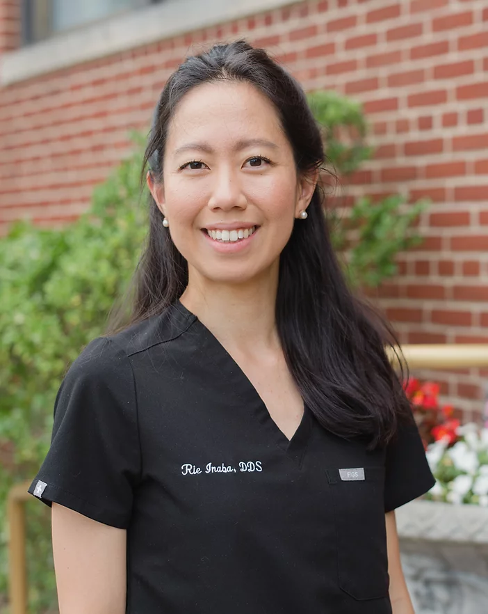
Rie Inaba, DDS
Dr. Rie Inaba, the eldest daughter of Dr. Takekazu Inaba, met her husband, Dr. Rhee, while attending dental school in California—despite having grown up just 30 minutes apart in New York. Dr. Inaba has extensive experience caring for individuals with intellectual and developmental disabilities, having previously worked in dental clinics specializing in these patient populations.
Outside of dentistry, Dr. Inaba and Dr. Rhee are proud parents to a daughter and a son, and enjoy spending time outdoors with their family—including their cat, Nori. Dr. Inaba is committed to providing family-oriented dentistry with an emphasis on patient comfort, clear communication, and high-quality care.
Education
- Loma Linda University School of Dentistry in California
- General Practice Residency at NYC Health + Hospital Bellevue
- Invisalign certified
Languages: English, Japanese
General Dentist
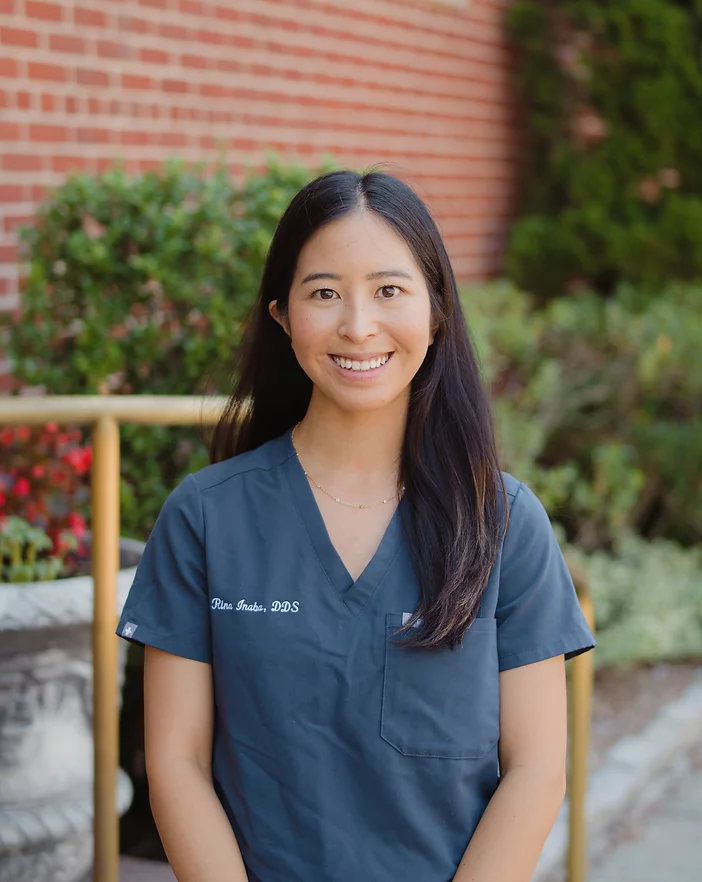
Rina Inaba, DDS
Dr. Rina Inaba is a graduate of Loma Linda University School of Dentistry, where she met her husband, Dr. Joshua Penners. After dental school, they moved to New York to complete a general dentistry residency training. Dr. Inaba is passionate about esthetic dentistry, with a focus on creating natural looking smiles while supporting long-term oral health. She is committed to providing high-quality, individualized care for every patient at Inaba Dental Office.
When she is not seeing patients, she is teaching at Touro College of Dental Medicine. Outside of dentistry, Dr. Inaba enjoys cooking, photography, and traveling!
Education
- Loma Linda University School of Dentistry in California
- General Practice Residency at St. Barnabas Hospital in Bronx
Languages: English, Japanese
General Dentist
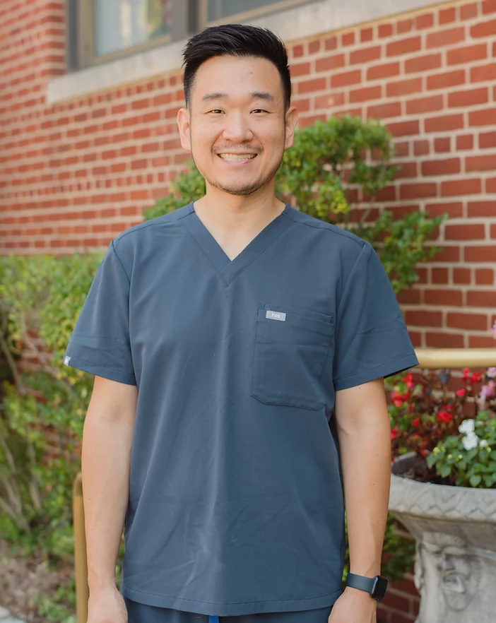
Matthew Rhee, DDS
Dr. Rhee and Dr. Rie Inaba are both New York natives who met while attending dental school in Loma Linda, California. Dr. Rhee has joined the team at Inaba Dental Office, where he brings advanced training from his residency and a commitment to delivering compassionate, high-quality care.
In addition to his clinical work, Dr. Rhee serves as a faculty member at Touro University College of Dental Medicine. He enjoys all aspects of general dentistry and has pursued additional training in root canal treatment.
Outside the office, Dr. Rhee and Dr. Inaba enjoy spending time outdoors and look forward to sharing their love of nature with their daughter and son as they grow.
Education
- Loma Linda University School of Dentistry in California
- General Practice Residency at the Veteran’s Administration Hospital in Brooklyn
- Dental implant surgical continuum at Implant Pathways
Languages: English, Korean
PGY2 Periodontics Resident
Joshua Penners, DDS
Dr. Joshua Penners was born and raised in California. As a California native, he loves the ocean and enjoys visiting national parks. He moved to New York after graduating from Loma Linda and completed a general practice residency program at the Brooklyn VA with an additional year at Bronx VA gaining more experience in surgical prosthodontic and periodontic procedures. After working full time in private practice, Dr. Penners decided to specialize in Periodontics and accepted a position at the Manhattan VA/NYU Periodontics training program. He hopes to provide comprehensive periodontal care that restores oral health in the context of the whole person.
Education
- Loma Linda University School of Dentistry in California
- General Practice Residency at the Veteran’s Administration Hospital in Brooklyn
- 2nd year General Practice Residency at the Veteran’s Administration Hospital in the Bronx
- Periodontics Resident at the Veteran’s Administration Hospital in Manhattan
Languages: English, Spanish
Office Manager
Miji Inaba
Miji Inaba started Inaba Dental Office with her husband, Dr. Takekazu Inaba. In addition to ensuring that all business aspects of the practice run smoothly and efficiently, Miji is responsible for filing insurance claims on patients’ behalf to help optimize their insurance benefits.
When she’s not at the office, Miji enjoys cooking and trying out new recipes, and stays busy with her grandchildren. She looks forward to welcoming you to Inaba Dental!
Languages: English, Japanese
Dental Assistant
Nami Colagrosso
Nami has been a part of the Inaba Dental team for over 20 years! Nami grew up in Mie, Japan, known for the Ise Grand Shrine, one of Japan’s holiest sites. She was a clinical instructor at a dental hygiene school in Japan before coming to New York. Nami enjoys reading and baking during her off time. Her favorite part about being at Inaba Dental is that she gets to be with the patients during every step of their treatment and help patients feel well cared for and comfortable.
Languages: English, Japanese
General Dentist
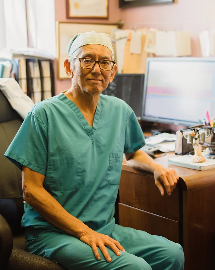
Takekazu Inaba, DDS
Dr. Takekazu Inaba went to Sophia University in Tokyo, Japan. He moved to California where he graduated from Loma Linda University in 1987. Dr. Inaba opened Inaba Dental Office at Scarsdale, NY in 1990 and at Westbury, NY in 2005. His daughters and sons-in-law continue the family dental practice.
Education
- Loma Linda University School of Dentistry in California
Languages: English, Japanese
Make an appointment.
Send an appointment request and our front desk will call you to schedule — or call either office to make an appointment.
Scarsdale Office
2 Overhill Road, Suite #110
Scarsdale, NY 10583
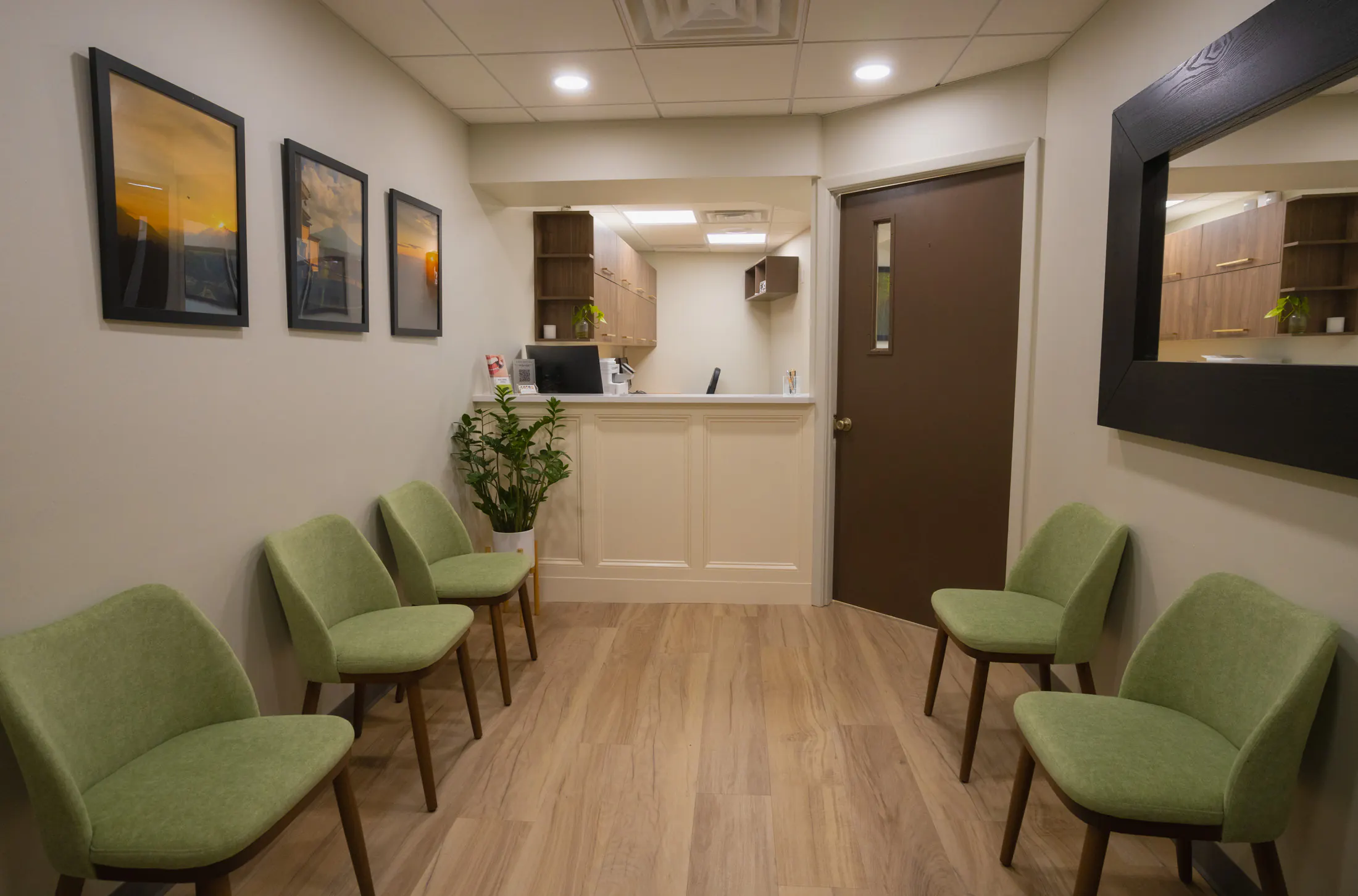
Westbury Office
265 Post Avenue, Suite #110
Westbury, NY 11590
PLACEHOLDER: Westbury interior photos, to be taken
Insurance and payment
Do you accept insurance?
Yes — we accept insurance, even though we don't participate as a "preferred provider" in your dental plan's network. Payment can happen in two ways: you pay for your services in full and your insurance company reimburses you — or you pay the remaining balance not included in your benefits after the claims have been processed.
We're happy to file insurance claims on your behalf, and we'll manage the paperwork and forms needed to optimize the reimbursement from your plan's coverage.
What is fee-for-service dentistry?
We are a fee-for-service dental practice, meaning we are not an in-network provider with dental insurance. This allows us to offer value-based care using high-quality materials, rather than have a third-party insurance provider decide what services and materials can be covered. It gives us as a provider — and you as a patient — more freedom in what's possible for your dental care.
Your family's dentist for as long as you're posted in New York.
Inaba Dental has cared for Japanese families in the New York area since 1990. Appointments are in Japanese. The front desk fills out your employer's health-insurance claim forms, and when you go home we prepare your records for your dentist in Japan.
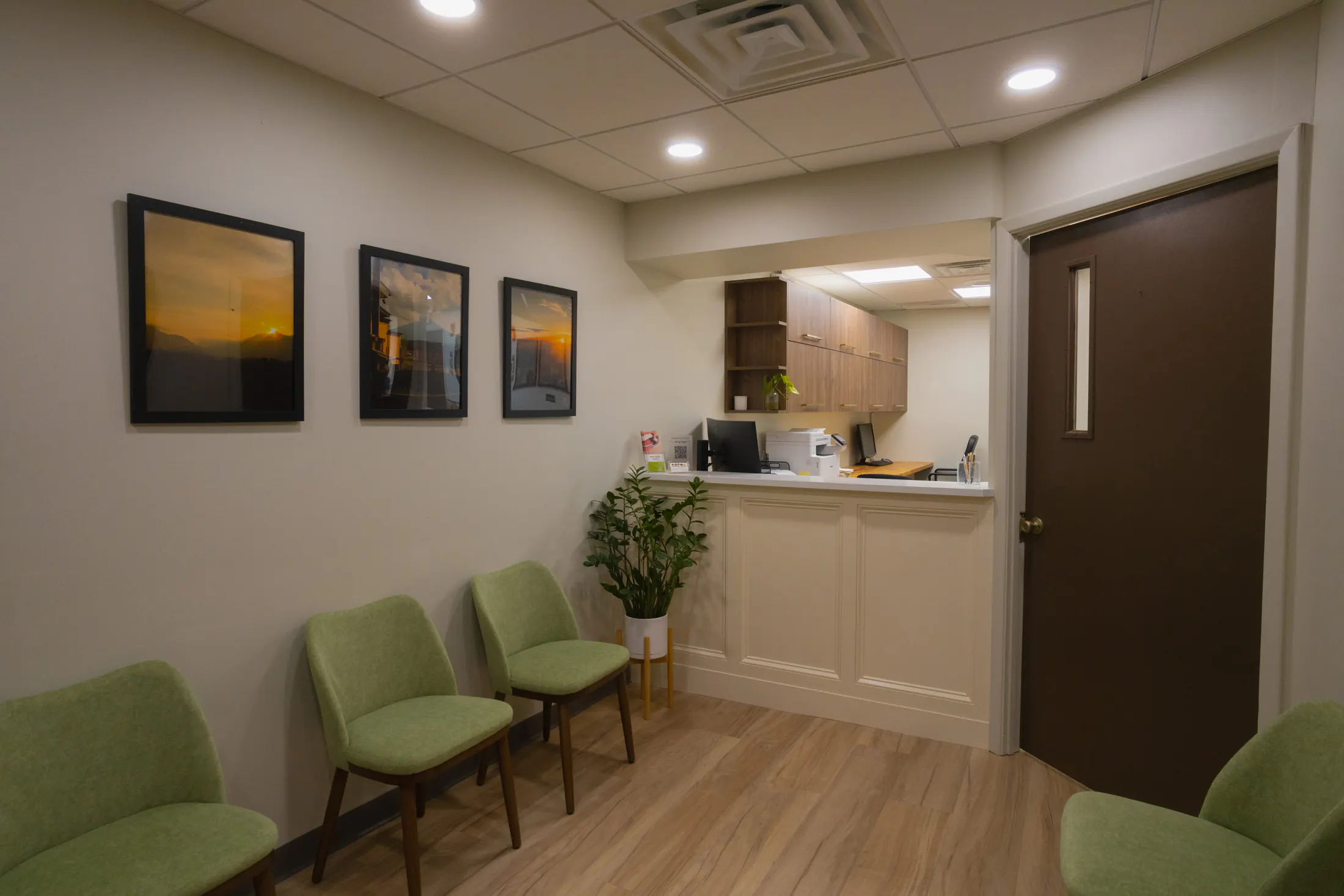
The front desk at our Scarsdale office — where your Japanese insurance paperwork is handled.
We fill out your Japanese insurance paperwork
If your employer reimburses dental care through a Japanese health-insurance plan, bring your company's claim forms. We complete the Japanese-language paperwork and support your Japan-side reimbursement.
We file your U.S. insurance claims
We are a fee-for-service practice and are not in any U.S. insurance network, which lets your doctor, not an insurance company, decide your treatment. We file claims with your U.S. plan for you. Out-of-network reimbursement usually arrives within two to four weeks, and we'll help you understand what your plan covers.
New York school dental forms
New York public schools require a dental certificate at enrollment. We perform the exam and complete the required school forms.
Moving home? Your records go with you
We prepare your treatment records and X-rays so your dentist in Japan can pick up right where we left off.
What to bring to your first visit
- Your dental insurance card (U.S. plan, if you have one)
- The policyholder's (世帯主) date of birth
- Your company's Japanese insurance claim forms, if your employer reimburses dental care
Japanese-speaking team
Dentist Dr. Rina Inaba
Dentist Miji Inaba
Front Desk & Insurance Nami Colagrosso
Clinical Assistant
Patient FAQ
Our patient FAQ pages cover these topics — currently written in Japanese.
PLACEHOLDER: English translations of the 9 Japanese FAQs (audit Action 9)
Send us a message
If you are having a dental emergency, please call us directly
Scarsdale 914.723.3511 or Westbury 516.242.9344
Scarsdale 914.723.3511 · Westbury 516.242.9344
What to bring to your first visit
We welcome new patients at both offices.
- Your insurance card
- The policyholder's date of birth
- Your company's claim forms, if your Japanese employer reimburses dental care
PLACEHOLDER: downloadable intake forms (English + Japanese PDFs)